What is venous ultrasound? Vein specialists use venous ultrasound to create images of the body veins. The ultrasound machine emits sound waves and creates vein images. Doctors use this diagnosis procedure to identify blood clots, especially in the leg veins. For instance, when you suffer from deep vein thrombosis, the doctor takes ultrasound images to identify the severity of the condition. Ultrasound is a safe diagnostic procedure because it doesn’t use ionizing radiation. These radiations are harmful to the body.

(Source)
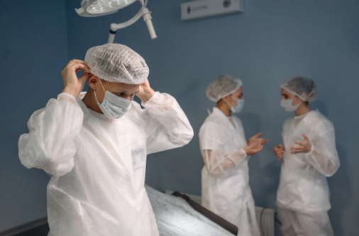
Before the procedure, the doctor may ask you to avoid drinking and eating for six to eight hours. Also, you should wear comfortable dresses and leave jewelry items at home. The doctor may also ask you to wear a gown.
Venous Ultrasound Imaging – What is it
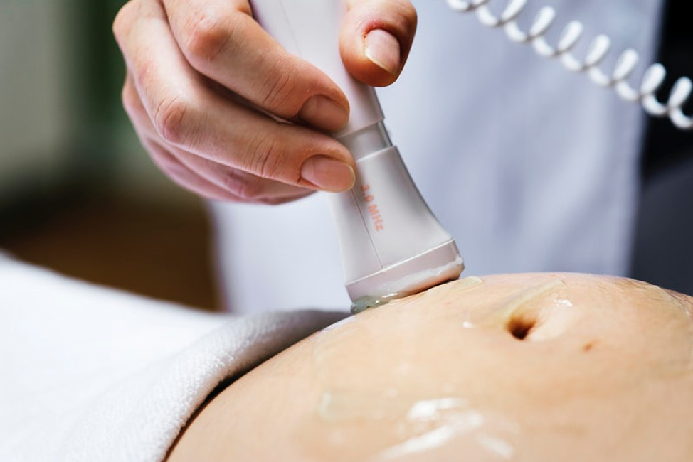
Ultrasound imaging helps vein specialists to diagnose venous and determine effective venous procedure. This is essentially a noninvasive medical procedure that helps doctors to produce pictures of veins through sound waves. The doctor applies gel on the skin and uses a transducer to take the image. The transducer is a small one that releases high-frequency sound waves. The probe collects the sound waves and produces an image on the computer.
The ultrasound machine creates images in real-time. This helps doctors to understand the vein structure. Also, doctors can view blood flow and spot the right position of the disorder. Venous ultrasound creates a picture of veins throughout the body.
The Common Uses of Venous Ultrasound
Vein specialists use ultrasound machines to identify blood clots, especially in the legs. Healthcare providers refer to this procedure as deep vein thrombosis. These blood clots travel throughout the body and block the blood supply to the lungs. These clots can cause life-threatening conditions such as pulmonary embolism. Upon early identification of blood clots, doctors can prevent the clot from passing to the lungs. The healthcare provider performs a venous ultrasound for the following reasons:
- It identifies long-standing leg swelling and varicose veins. When the valves inside the veins are damaged, the blood pools in the vein. The venous ultrasound identifies the blood pool due to varicose veins. Doctors can identify the problem and remove the veins from the body.
- Venous ultrasound helps in performing treatment efficiently. While performing invasive procedures, the doctor can check the right position to enter needles and catheters. This helps doctors to prevent bleeding and nerve damage.
- Healthcare professionals can mark the veins and arteries before performing a procedure. Also, they can identify whether it is safe to perform the procedure or there are complications. For instance, when doctors remove the superficial varicose veins, they should assure the right location for incision.
- They can assess a blood vessel graft for dialysis, especially when the graft is blocked or narrowed.
The doctor might also use venous ultrasound in children. During congenital vascular malformation and dialysis fistula, the doctor may use venous ultrasound. It helps them identify the connection between a vein and an artery.
While performing a surgery, the doctor places a tiny incision on the children’s leg. This might cause blood clotting due to small vessel size. Also, a blood clot might appear on the arm due to compression. In both conditions, the doctor uses venous ultrasound to evaluate the arteries. Here are some benefits of using venous ultrasound for doctors:
- Identification of blood clotting
- Increase in blood flow due to infection
- Reduced blood flow to various organs
- Tumor and congenital vascular malformation
- Narrowing of vessels

(Source)
Things to Remember Before the Procedure
Before the procedure you should:
- Consume every medication prescribed by the doctor
- Wear comfortable clothes
- Arrive 15 minutes before the appointment
During Your Exam
When you arrive at the doctor’s clinic, they will ask you to:
- Lie on the examination table with your hands on the side. The doctor will apply a gel and place the transducer on the veins and arteries they want to capture.
- When the physician will perform the diagnosis, you might hear the blood pumping throughout the room. You don’t need to be afraid as this is normal.
- The examination might take up to 15 to 30 minutes.
After Your Exam
After the examination, you can walk and even leave for your home. There isn’t any post-exam instruction. A vein specialist will review your results and identify your health condition.
Venous Ultrasound: Conclusion
A venous ultrasound is a simple and harmless procedure that doesn’t include any side effects. The device uses sound waves to captures the veins. With the help of this diagnosis, vein specialists can identify varicose veins and other venous conditions. Furthermore, they can also spot blood clots and performs procedures to prevent life-threatening conditions.
Do you live in New York and want to consult a vein specialist? If yes, then you should contact the Vascular Surgery and Vein Center in New York. Our experienced vein specialist, Dr. Norman Chideckel treats a wide range of venous diseases. He uses advanced procedures, both non-invasive and invasive to treat conditions such as varicose veins.
For more information, give us a call at 212-993-6133.


